개요 (Overview)
망막모세포종(Retinoblastoma)은 소아에서 가장 흔한 안내 악성종양입니다. RB1 종양억제 유전자 변이에 의해 발생하며, **백색동공(leukocoria)**이 가장 흔한 발현 양상입니다. 유전성(양안, 40%)과 비유전성(편측, 60%)으로 나뉩니다.
Knudson's Two-hit Hypothesis
- 유전성: 1st hit = 생식세포 변이 (모든 세포). 2nd hit = 체세포 변이 → 양안, 조기, 다발
- 비유전성: 1st + 2nd hit 모두 체세포 → 편측, 단발, 늦은 발병
임상 소견
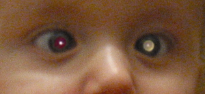
- Leukocoria (백색동공): 가장 흔한 발현 (60%)
- 사시: 두 번째 (20%)
- 안구 내 석회화: B-scan/CT에서 특징적
- 성장 패턴: endophytic (유리체 내), exophytic (망막하), diffuse (평평)
시험 포인트 / Board Points
Board Point
- Leukocoria 감별의 1순위 = retinoblastoma
- RB1 유전자 (13q14) — 종양억제 유전자
- Knudson's two-hit hypothesis
- 석회화 = retinoblastoma의 특징 (Coats, PFV에는 없음)
- MRI 선호 (CT는 방사선 → 2차 종양 위험 증가)
- Trilateral Rb: 양안 Rb + pinealoblastoma
- Flexner-Wintersteiner rosette: 특징적 조직소견
- 적출 시 시신경 절단연 확인 (시신경 침범 = 예후 불량)
Warning
Retinoblastoma에서 조직검사(biopsy)는 금기! 종양 파종 위험. 임상+영상으로 진단. 적출 후 조직 확인.
Guideline Snapshot— ICRB / COG
- ICRB Group A-E로 분류
- Group A-B: focal therapy ± chemoreduction
- Group C-D: IAC or systemic chemo
- Group E: 적출 고려
- 유전성: 유전 상담 + 형제/자녀 선별
참고문헌 (References)
- Shields CL, Shields JA. Retinoblastoma management: advances in enucleation, intravenous chemoreduction, and intra-arterial chemotherapy. Curr Opin Ophthalmol. 2010;21(3):203-212.
- Dimaras H, Kimani K, et al. Retinoblastoma. Lancet. 2012;379(9824):1436-1446.