개요 (Overview)
맥락막골종(Choroidal Osteoma)은 맥락막 내에 성숙 골 조직이 형성되는 드문 양성 종양입니다. 젊은 여성에서 호발하며, **시신경유두 주위(juxtapapillary)**에 주황색/황백색의 편평한 종괴로 나타납니다. CT에서 골밀도가 확인되면 진단적입니다.
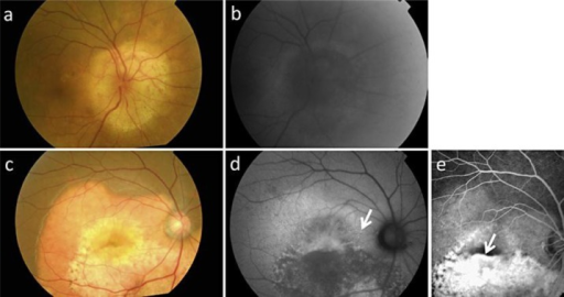
임상 소견
- 주황색/황백색 편평 종괴: 시신경유두 주위
- 경계 명확, 과결절/과쪽으로 확장 가능
- B-scan: 고반사 + 후방음영(posterior shadowing) = 특징적
- CT: 맥락막 내 골밀도(bony density) = 진단적
- OCT-EDI: 맥락막 내 고반사 층
시험 포인트 / Board Points
Board Point
- 젊은 여성 호발
- 시신경유두 주위(juxtapapillary) 편평 종괴
- B-scan: 고반사 + 후방음영 (bone)
- CT: 골밀도 = 진단적
- 양성 — 관찰이 기본
- CNV: 시력저하의 주 원인 → anti-VEGF
- 탈석회화(decalcification): RPE 위축, 비가역적 시력 저하
Exam Point
맥락막 종괴에서 후방음영(posterior shadowing) on B-scan = 석회화 시사 → 맥락막골종 또는 망막모세포종(소아). CT에서 골밀도 확인으로 감별.
Guideline Snapshot— Expert consensus
- CT에서 골밀도 확인 = 진단
- CNV 없으면 관찰
- CNV → anti-VEGF
- 장기 추적 (탈석회화, CNV 감시)
참고문헌 (References)
- Shields CL, Sun H, et al. Choroidal osteoma: clinical features, mushroom configuration, and treatment in 74 patients. Ophthalmology. 2005;112(10):1709-1720.
- Aylward GW, Chang TS, et al. Choroidal osteoma and choroidal neovascularization. Br J Ophthalmol. 1998;82(5):588-599.