개요 (Overview)
악센펠트-리거증후군(Axenfeld-Rieger Syndrome, ARS)은 신경능선세포(neural crest cell)의 이동/분화 이상으로 전방각, 홍채, 각막에 발달 이상이 발생하고, 전신 이상(치아, 안면, 배꼽)이 동반되는 상염색체 우성 유전 질환입니다.
약 **50%**에서 녹내장이 발생합니다.
스펙트럼 분류
| 명칭 | 소견 |
|---|---|
| Axenfeld anomaly | Posterior embryotoxon + 전방각에 iris strands |
| Rieger anomaly | Axenfeld anomaly + 홍채 이상 (corectopia, polycoria, 홍채 위축) |
| Rieger syndrome | Rieger anomaly + 전신 이상 (치아, 안면, 배꼽) |
| Axenfeld-Rieger syndrome | 이들의 통합 용어 |
역학 (Epidemiology)
- 약 1:200,000 출생
- 상염색체 우성 — 가변적 표현도(variable expressivity)
- 양안성
- 녹내장 발생률: 약 50% (소아~청소년기)
원인/병태생리 (Pathophysiology)
- PITX2 (4q25) 또는 FOXC1 (6p25) 유전자 돌연변이
- 신경능선세포 유래 조직의 발달 이상
- 전방각 이상 → 방수 유출 장애 → 녹내장
Exam Point
Posterior embryotoxon은 Schwalbe line의 전방 변위 및 비후로, 정상 인구의 약 8-15%에서도 관찰됩니다. 그러나 홍채 이상이 동반되면 ARS를 의심해야 합니다.
분류 및 증상/징후 (Classification & Signs)
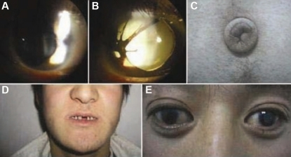
안과적 소견
- Posterior embryotoxon: Schwalbe line의 전방 변위 — 세극등에서 각막 주변부에 흰색 선
- Iris strands: 홍채에서 posterior embryotoxon으로 연결되는 조직 가닥 (전방각경)
- 홍채 이상: Corectopia (동공 편위), polycoria, iris atrophy
- 녹내장: 약 50%
전신 이상 (Rieger syndrome)
- 치아: Microdontia, hypodontia, oligodontia
- 안면: Maxillary hypoplasia, telecanthus, 편평한 mid-face
- 배꼽: Periumbilical skin redundancy
Board Point
ARS의 안과적 소견 3단계:
- Posterior embryotoxon (Schwalbe line 전방 변위)
-
- Iris strands (Axenfeld anomaly)
-
- 홍채 이상: corectopia, polycoria (Rieger anomaly)
-
- 전신이상: 치아, 안면, 배꼽 (Rieger syndrome)
진단 (Diagnosis)
- 세극등: Posterior embryotoxon, 홍채 이상
- 전방각경: Iris strands, 전방각 이상
- 전신검사: 치아, 안면, 배꼽
- 유전자검사: PITX2, FOXC1
치료 (Treatment)
- 약물: POAG와 동일
- 수술: Goniotomy/trabeculotomy (효과 제한적일 수 있음) → GDD
- 전신이상에 대한 다학제 관리
예후/경과 (Prognosis)
- 50%에서 녹내장 발생 — 소아~청소년기에 주로 시작
- 수술적 치료가 필요한 경우 많음
- 약시 동반 가능 → 조기 치료
시험 포인트 / Board Points
Board Point
- Posterior embryotoxon ⭐⭐⭐: 가장 특징적 소견
- 전신 이상 (치아, 안면, 배꼽) ⭐⭐⭐: Rieger syndrome
- 50% 녹내장 ⭐⭐
- PITX2, FOXC1 ⭐⭐: 상염색체 우성
- 양안성: ICE(편측)와 감별
- Posterior embryotoxon 단독: 정상 변이(8-15%)일 수 있음
Controversies / Recent Updates
- 유전자형-표현형 상관관계: PITX2 vs FOXC1에 따른 표현형 차이 연구
Guideline Snapshot— AAPOS / Expert Consensus
- ARS 진단 시 전신 검사 필수 (치아, 안면, 배꼽)
- 안압 정기 추적 (50% 녹내장)
- 가족 선별검사 (상염색체 우성)
- 녹내장 치료: 약물 → 수술
참고문헌 (References)
- Tumer Z, Bach-Holm D. Axenfeld-Rieger syndrome and spectrum of PITX2 and FOXC1 mutations. Eur J Hum Genet. 2009;17(12):1527-1539.
- Shields MB, Buckley E, et al. Axenfeld-Rieger syndrome: a spectrum of developmental disorders. Surv Ophthalmol. 1985;29(6):387-409.